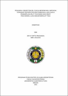
Pengaruh Sekretom Sel Punca Mesenkimal Hipoksia terhadap Percepatan Penyembuhan Luka Pasca Penempelan Split Thickness Skin Graft pada Model Luka Bakar Derajat Tiga

Date
2023Author
Prananda, Arya Tjipta
Advisor(s)
Ilyas, Syafruddin
Putra, Agung
Nasution, Iqbal Pahlevi Adeputra
Metadata
Show full item recordAbstract
Background: Effective wound management is an important determinant of survival and prognosis of third-degree burn patients. Recently, autologous skin grafting techniques (skin grafts) such as split-thickness grafts (STSG) are still the standard therapy in the treatment of third-degree burns. The effectiveness of STSG is still low due to the limitations of skin donors, especially in patients with third degree burns. Due to the limitations of this therapy, patients experience grafting failure of about 5-10%, delayed wound closure, infection, appearance of scar tissue, and even death. Therefore, new techniques for full and timely burn closure are urgently needed. Recent research has shown that sekretom mesenchymal stem cells (H-MSCs) contain bioactive soluble molecules such as growth factors and anti-inflammatory cytokines that are able to control inflammation which has an impact on improving skin function and structure. However, the benefits of using sekretom H-MSCs in the repair of third-degree burns are still unclear.
Objective: The purpose of this study was to determine the effect of using sekretom H-MSCs on accelerating wound healing after STSG attachment in a rat model of third-degree burns in vivo.
Methods: This study is an experimental laboratory in vivo post-test only control group design with 5 treatment groups consisting of sham, control, P1 (split-thickness grafts), P2 (split-thickness grafts and sekretom H-MSCs), and P3 (sekretom H-MSCs). The STSG technique is performed by attaching 1.5 cm of thigh skin to the burn area 24 hours after burn induction. Sekretom H-MSCs 200 l were administered topically on the STSG section. Measurement of the degree of inflammation by measuring the expression of IL-1, IL-10, TNF-α, and PCNA with western blot. Analysis of the proliferation process by observing angiogenesis with evaluation of VEGF levels by ELISA. Analysis of the remodeling process through the analysis of collagen with specific staining of Masson trichome. Data analysis was performed by descriptive linear regression and one-way ANOVA.
Results: Application of sekretom H-MSCs (P2) 200 μL increased the amount of epithelialization, increased the percentage of vascularization, increased the number of keratinocytes, increased IL-10 protein expression, decreased IL-1β protein expression, and decreased IL-6 gene expression, increased fibronectin gene expression, increased PCNA protein expression, increased VEGF gene expression, increased α-SMA protein expression, and increased the amount of collagen post split thickness skin graft in third degree burn models when compared to controls.
Conclusion: Application of sekretom H-MSCs accelerated the healing of third degree burns after split thickness skin graft in third degree burn rats.
Collections
- Doctoral Dissertations [194]
